Artlabeling Activity Comparing the Somatic Motor and Autonomic Innervation
| Autonomic nervous arrangement | |
|---|---|
 Autonomic nervous system innervation. | |
| Details | |
| Identifiers | |
| Latin | Autonomici systematis nervosi |
| MeSH | D001341 |
| TA98 | A14.3.00.001 |
| TA2 | 6600 |
| FMA | 9905 |
| Anatomical terminology [edit on Wikidata] | |
The autonomic nervous system (ANS), formerly referred to as the vegetative nervous system, is a division of the peripheral nervous system that supplies polish muscle and glands, and thus influences the function of internal organs.[1] The autonomic nervous system is a control system that acts largely unconsciously and regulates bodily functions, such as the heart rate, digestion, respiratory rate, pupillary response, urination, and sexual arousal.[2] This system is the main machinery in command of the fight-or-flying response.
The autonomic nervous organization is regulated past integrated reflexes through the brainstem to the spinal cord and organs. Autonomic functions include command of respiration, cardiac regulation (the cardiac control center), vasomotor activity (the vasomotor middle), and certain reflex deportment such as coughing, sneezing, swallowing and vomiting. Those are and so subdivided into other areas and are also linked to autonomic subsystems and the peripheral nervous organization. The hypothalamus, but in a higher place the brain stem, acts every bit an integrator for autonomic functions, receiving autonomic regulatory input from the limbic organization.[3]
The autonomic nervous system has three branches: the sympathetic nervous organization, the parasympathetic nervous system and the enteric nervous organisation.[4] [v] [6] [7] Some textbooks do not include the enteric nervous organisation equally part of this system.[eight] The sympathetic nervous organization is oftentimes considered the "fight or flight" organization, while the parasympathetic nervous system is often considered the "balance and digest" or "feed and breed" system. In many cases, both of these systems accept "opposite" actions where one arrangement activates a physiological response and the other inhibits it. An older simplification of the sympathetic and parasympathetic nervous systems as "excitatory" and "inhibitory" was overturned due to the many exceptions plant. A more modernistic characterization is that the sympathetic nervous system is a "quick response mobilizing system" and the parasympathetic is a "more than slowly activated dampening system", just even this has exceptions, such as in sexual arousal and orgasm, wherein both play a function.[3]
There are inhibitory and excitatory synapses between neurons. A third subsystem of neurons has been named equally non-noradrenergic, non-cholinergic transmitters (considering they employ nitric oxide as a neurotransmitter) and are integral in autonomic role, in particular in the gut and the lungs.[9]
Although the ANS is besides known equally the visceral nervous system, the ANS is merely connected with the motor side.[10] Nearly autonomous functions are involuntary but they tin often work in conjunction with the somatic nervous system which provides voluntary control.
Structure [edit]
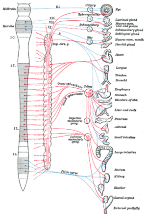
Autonomic nervous system, showing splanchnic nerves in heart, and the vagus nervus as "10" in blue. The heart and organs beneath in list to right are regarded equally viscera.
The autonomic nervous organization is divided into the sympathetic nervous system and parasympathetic nervous system. The sympathetic division emerges from the spinal cord in the thoracic and lumbar areas, terminating effectually L2-3. The parasympathetic partitioning has craniosacral "outflow", pregnant that the neurons brainstorm at the cranial nerves (specifically the oculomotor nerve, facial nervus, glossopharyngeal nervus and vagus nerve) and sacral (S2-S4) spinal cord.
The autonomic nervous organisation is unique in that it requires a sequential two-neuron efferent pathway; the preganglionic neuron must first synapse onto a postganglionic neuron earlier innervating the target organ. The preganglionic, or beginning, neuron will begin at the "outflow" and volition synapse at the postganglionic, or 2d, neuron'southward jail cell torso. The postganglionic neuron volition so synapse at the target organ.
Sympathetic partition [edit]
The sympathetic nervous system consists of cells with bodies in the lateral grey column from T1 to L2/3. These cell bodies are "GVE" (general visceral efferent) neurons and are the preganglionic neurons. In that location are several locations upon which preganglionic neurons can synapse for their postganglionic neurons:
- Paravertebral ganglia (3) of the sympathetic chain (these run on either side of the vertebral bodies)
- cervical ganglia (3)
- thoracic ganglia (12) and rostral lumbar ganglia (2 or iii)
- caudal lumbar ganglia and sacral ganglia
- Prevertebral ganglia (celiac ganglion, aorticorenal ganglion, superior mesenteric ganglion, inferior mesenteric ganglion)
- Chromaffin cells of the adrenal medulla (this is the one exception to the ii-neuron pathway rule: the synapse is direct efferent onto the target jail cell bodies)
These ganglia provide the postganglionic neurons from which innervation of target organs follows. Examples of splanchnic (visceral) nerves are:
- Cervical cardiac nerves and thoracic visceral nerves, which synapse in the sympathetic chain
- Thoracic splanchnic fretfulness (greater, lesser, to the lowest degree), which synapse in the prevertebral ganglia
- Lumbar splanchnic nerves, which synapse in the prevertebral ganglia
- Sacral splanchnic fretfulness, which synapse in the inferior hypogastric plexus
These all contain afferent (sensory) nerves too, known equally GVA (general visceral afferent) neurons.
Parasympathetic division [edit]
The parasympathetic nervous system consists of cells with bodies in one of two locations: the brainstem (Cranial Nerves 3, Seven, IX, X) or the sacral spinal cord (S2, S3, S4). These are the preganglionic neurons, which synapse with postganglionic neurons in these locations:
- Parasympathetic ganglia of the head: Ciliary (Cranial nerve III), Submandibular (Cranial nervus Vii), Pterygopalatine (Cranial nerve 7), and Otic (Cranial nerve IX)
- In or near the wall of an organ innervated by the Vagus (Cranial nerve X) or Sacral nerves (S2, S3, S4)
These ganglia provide the postganglionic neurons from which innervations of target organs follows. Examples are:
- The postganglionic parasympathetic splanchnic (visceral) fretfulness
- The vagus nerve, which passes through the thorax and abdominal regions innervating, among other organs, the heart, lungs, liver and stomach
Sensory neurons [edit]
The sensory arm is equanimous of main visceral sensory neurons found in the peripheral nervous system (PNS), in cranial sensory ganglia: the geniculate, petrosal and nodose ganglia, appended respectively to cranial nerves VII, IX and Ten. These sensory neurons monitor the levels of carbon dioxide, oxygen and saccharide in the blood, arterial force per unit area and the chemical limerick of the tummy and gut content. They too convey the sense of taste and smell, which, unlike about functions of the ANS, is a conscious perception. Blood oxygen and carbon dioxide are in fact direct sensed by the carotid body, a pocket-sized drove of chemosensors at the bifurcation of the carotid artery, innervated by the petrosal (IXth) ganglion. Main sensory neurons project (synapse) onto "2nd order" visceral sensory neurons located in the medulla oblongata, forming the nucleus of the alone tract (nTS), that integrates all visceral information. The nTS as well receives input from a nearby chemosensory center, the surface area postrema, that detects toxins in the claret and the cerebrospinal fluid and is essential for chemically induced vomiting or provisional taste aversion (the memory that ensures that an animal that has been poisoned past a food never touches information technology over again). All this visceral sensory information constantly and unconsciously modulates the activeness of the motor neurons of the ANS.
Innervation [edit]
Autonomic fretfulness travel to organs throughout the trunk. Most organs receive parasympathetic supply past the vagus nerve and sympathetic supply by splanchnic fretfulness. The sensory part of the latter reaches the spinal column at certain spinal segments. Pain in whatever internal organ is perceived as referred pain, more specifically as pain from the dermatome corresponding to the spinal segment.[11]
| Organ | Fretfulness[12] | Spinal column origin[12] |
|---|---|---|
| stomach |
| T5, T6, T7, T8, T9, sometimes T10 |
| duodenum |
| T5, T6, T7, T8, T9, sometimes T10 |
| jejunum and ileum |
| T5, T6, T7, T8, T9 |
| spleen |
| T6, T7, T8 |
| gallbladder and liver |
| T6, T7, T8, T9 |
| colon |
|
|
| pancreatic caput |
| T8, T9 |
| appendix |
| T10 |
| kidneys and ureters |
| T11, T12 |
Motor neurons [edit]
Motor neurons of the autonomic nervous organisation are institute in ''autonomic ganglia''. Those of the parasympathetic branch are located shut to the target organ whilst the ganglia of the sympathetic branch are located close to the spinal string.
The sympathetic ganglia here, are constitute in two chains: the pre-vertebral and pre-aortic bondage. The activity of autonomic ganglionic neurons is modulated by "preganglionic neurons" located in the central nervous organisation. Preganglionic sympathetic neurons are located in the spinal cord, at the thorax and upper lumbar levels. Preganglionic parasympathetic neurons are found in the medulla oblongata where they grade visceral motor nuclei; the dorsal motor nucleus of the vagus nervus; the nucleus ambiguus, the salivatory nuclei, and in the sacral region of the spinal cord.
Role [edit]
Function of the autonomic nervous arrangement [13]
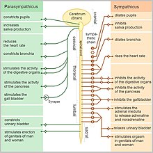
Sympathetic and parasympathetic divisions typically function in opposition to each other. Just this opposition is better termed complementary in nature rather than antagonistic. For an analogy, i may remember of the sympathetic division equally the accelerator and the parasympathetic sectionalisation as the brake. The sympathetic sectionalization typically functions in deportment requiring quick responses. The parasympathetic segmentation functions with deportment that do not require immediate reaction. The sympathetic organisation is often considered the "fight or flight" organisation, while the parasympathetic system is oftentimes considered the "rest and digest" or "feed and breed" system.
Nonetheless, many instances of sympathetic and parasympathetic activity cannot be ascribed to "fight" or "rest" situations. For example, standing up from a reclining or sitting position would entail an unsustainable drop in claret force per unit area if not for a compensatory increment in the arterial sympathetic tonus. Some other example is the constant, 2nd-to-2nd, modulation of heart rate by sympathetic and parasympathetic influences, as a role of the respiratory cycles. In general, these ii systems should exist seen as permanently modulating vital functions, in a usually antagonistic fashion, to accomplish homeostasis. Higher organisms maintain their integrity via homeostasis which relies on negative feedback regulation which, in plough, typically depends on the autonomic nervous organisation.[14] Some typical deportment of the sympathetic and parasympathetic nervous systems are listed below.[15]
| Target organ/system | Parasympathetic | Sympathetic |
|---|---|---|
| Digestive system | Increase peristalsis and corporeality of secretion past digestive glands | Decrease activity of digestive system |
| Liver | No result | Causes glucose to exist released to blood |
| Lungs | Constricts bronchioles | Dilates bronchioles |
| Urinary bladder/ Urethra | Relaxes sphincter | Constricts sphincter |
| Kidneys | No effects | Decrease urine output |
| Center | Decreases rate | Increase rate |
| Claret vessels | No effect on nigh blood vessels | Constricts claret vessels in viscera; increase BP |
| Salivary and Lacrimal glands | Stimulates; increases product of saliva and tears | Inhibits; result in dry mouth and dry eyes |
| Eye (iris) | Stimulates constrictor muscles; constrict pupils | Stimulate dilator muscle; dilates pupils |
| Eye (ciliary muscles) | Stimulates to increase jutting of lens for shut vision | Inhibits; decrease bulging of lens; prepares for afar vision |
| Adrenal Medulla | No effect | Stimulate medulla cells to secrete epinephrine and norepinephrine |
| Sweat gland of peel | No issue | Stimulate sudomotor role to produce perspiration |
Sympathetic nervous system [edit]
Promotes a fight-or-flight response, corresponds with arousal and energy generation, and inhibits digestion
- Diverts blood flow away from the gastro-intestinal (GI) tract and skin via vasoconstriction
- Blood menstruation to skeletal muscles and the lungs is enhanced (by as much as 1200% in the example of skeletal muscles)
- Dilates bronchioles of the lung through circulating epinephrine, which allows for greater alveolar oxygen exchange
- Increases centre rate and the contractility of cardiac cells (myocytes), thereby providing a mechanism for enhanced blood catamenia to skeletal muscles
- Dilates pupils and relaxes the ciliary muscle to the lens, allowing more than light to enter the eye and enhances far vision
- Provides vasodilation for the coronary vessels of the center
- Constricts all the intestinal sphincters and the urinary sphincter
- Inhibits peristalsis
- Stimulates orgasm
The pattern of innervation of the sweat gland—namely, the postganglionic sympathetic nervus fibers—allows clinicians and researchers to use sudomotor function testing to assess dysfunction of the autonomic nervous systems, through electrochemical pare conductance.
Parasympathetic nervous organization [edit]
The parasympathetic nervous organisation has been said to promote a "rest and assimilate" response, promotes calming of the nerves render to regular function, and enhancing digestion. Functions of fretfulness within the parasympathetic nervous system include:[ citation needed ]
- Dilating blood vessels leading to the GI tract, increasing the claret flow.
- Constricting the bronchiolar diameter when the need for oxygen has diminished
- Dedicated cardiac branches of the vagus and thoracic spinal accessory fretfulness impart parasympathetic command of the heart (myocardium)
- Constriction of the pupil and contraction of the ciliary muscles, facilitating adaptation and allowing for closer vision
- Stimulating salivary gland secretion, and accelerates peristalsis, mediating digestion of food and, indirectly, the assimilation of nutrients
- Sexual. Nerves of the peripheral nervous arrangement are involved in the erection of genital tissues via the pelvic splanchnic nerves 2–iv. They are likewise responsible for stimulating sexual arousal.
Enteric nervous arrangement [edit]
The enteric nervous system is the intrinsic nervous system of the gastrointestinal organisation. It has been described as "the Second Brain of the Man Body".[xvi] Its functions include:
- Sensing chemical and mechanical changes in the gut
- Regulating secretions in the gut
- Controlling peristalsis and some other movements
Neurotransmitters [edit]

A flow diagram showing the process of stimulation of adrenal medulla that makes information technology release adrenaline, that further acts on adrenoreceptors, indirectly mediating or mimicking sympathetic action.

At the effector organs, sympathetic ganglionic neurons release noradrenaline (norepinephrine), along with other cotransmitters such every bit ATP, to act on adrenergic receptors, with the exception of the sweat glands and the adrenal medulla:
- Acetylcholine is the preganglionic neurotransmitter for both divisions of the ANS, as well as the postganglionic neurotransmitter of parasympathetic neurons. Nerves that release acetylcholine are said to be cholinergic. In the parasympathetic organization, ganglionic neurons use acetylcholine as a neurotransmitter to stimulate muscarinic receptors.
- At the adrenal medulla, there is no postsynaptic neuron. Instead, the presynaptic neuron releases acetylcholine to act on nicotinic receptors. Stimulation of the adrenal medulla releases adrenaline (epinephrine) into the bloodstream, which acts on adrenoceptors, thereby indirectly mediating or mimicking sympathetic activity.
A full table is found at Tabular array of neurotransmitter actions in the ANS.
History [edit]
The specialised organization of the autonomic nervous system was recognised by Galen. In 1665, Willis used the terminology, and in 1900, Langley used the term, defining the two divisions as the sympathetic and parasympathetic nervous systems.[17]
Caffeine effects [edit]
Caffeine is a bioactive ingredient constitute in commonly consumed beverages such as java, tea, and sodas. Brusk-term physiological furnishings of caffeine include increased claret force per unit area and sympathetic nerve outflow. Habitual consumption of caffeine may inhibit physiological brusque-term furnishings. Consumption of caffeinated espresso increases parasympathetic activity in habitual caffeine consumers; however, decaffeinated espresso inhibits parasympathetic activity in habitual caffeine consumers. Information technology is possible that other bioactive ingredients in decaffeinated espresso may likewise contribute to the inhibition of parasympathetic activity in habitual caffeine consumers.[xviii]
Caffeine is capable of increasing work capacity while individuals perform strenuous tasks. In one study, caffeine provoked a greater maximum heart rate while a strenuous task was being performed compared to a placebo. This trend is likely due to caffeine'southward ability to increase sympathetic nerve outflow. Furthermore, this study found that recovery later intense practice was slower when caffeine was consumed prior to exercise. This finding is indicative of caffeine's trend to inhibit parasympathetic activity in non-habitual consumers. The caffeine-stimulated increase in nerve activity is likely to evoke other physiological effects as the trunk attempts to maintain homeostasis.[nineteen]
The furnishings of caffeine on parasympathetic activity may vary depending on the position of the individual when autonomic responses are measured. One study found that the seated position inhibited autonomic action afterward caffeine consumption (75 mg); withal, parasympathetic activeness increased in the supine position. This finding may explain why some habitual caffeine consumers (75 mg or less) do not experience brusk-term furnishings of caffeine if their routine requires many hours in a seated position. It is important to note that the data supporting increased parasympathetic activeness in the supine position was derived from an experiment involving participants between the ages of 25 and 30 who were considered healthy and sedentary. Caffeine may influence autonomic activity differently for individuals who are more active or elderly.[20]
Come across likewise [edit]
- Dysautonomia
- International Gild for Autonomic Neuroscience
- Medullary ischemic reflex
References [edit]
- ^ "autonomic nervous system" at Dorland's Medical Dictionary
- ^ Schmidt, A; Thews, Yard (1989). "Autonomic Nervous System". In Janig, Westward (ed.). Human Physiology (2 ed.). New York, NY: Springer-Verlag. pp. 333–370.
- ^ a b Allostatic load notebook: Parasympathetic Office Archived 2012-08-19 at the Wayback Machine - 1999, MacArthur research network, UCSF
- ^ Langley, J.N. (1921). The Autonomic Nervous System Part 1. Cambridge: W. Heffer.
- ^ Jänig, Wilfrid (2008). Integrative action of the autonomic nervous system : neurobiology of homeostasis (Digitally printed version. ed.). Cambridge: Cambridge University Press. p. 13. ISBN978052106754-6.
- ^ Furness, John (nine October 2007). "Enteric nervous system". Scholarpedia. ii (10): 4064. Bibcode:2007SchpJ...2.4064F. doi:10.4249/scholarpedia.4064.
- ^ Willis, William D. (2004). "The Autonomic Nervous System and its key command". In Berne, Robert Yard. (ed.). Physiology (5. ed.). St. Louis, Mo.: Mosby. ISBN0323022251.
- ^ Pocock, Gillian (2006). Man Physiology (tertiary ed.). Oxford University Press. pp. 63–64. ISBN978-0-19-856878-0.
- ^ Belvisi, Maria Thou.; David Stretton, C.; Yacoub, Magdi; Barnes, Peter J. (1992). "Nitric oxide is the endogenous neurotransmitter of bronchodilator nerves in humans". European Journal of Pharmacology. 210 (2): 221–2. doi:10.1016/0014-2999(92)90676-U. PMID 1350993.
- ^ Costanzo, Linda S. (2007). Physiology . Hagerstwon, MD: Lippincott Williams & Wilkins. p. 37. ISBN978-0-7817-7311-9.
- ^ Essential Clinical Anatomy. K.L. Moore & A.One thousand. Agur. Lippincott, two ed. 2002. Folio 199
- ^ a b Unless specified otherwise in the boxes, the source is: Moore, Keith L.; Agur, A. M. R. (2002). Essential Clinical Anatomy (2nd ed.). Lippincott Williams & Wilkins. p. 199. ISBN978-0-7817-5940-three.
- ^ Neil A. Campbell, Jane B. Reece: Biologie. Spektrum-Verlag Heidelberg-Berlin 2003, ISBN 3-8274-1352-four
- ^ Goldstein, David (2016). Principles of Autonomic Medicine (PDF) (free online version ed.). Bethesda, Maryland: National Plant of Neurological Disorders and Stroke, National Institutes of Health. ISBN9780824704087. Archived from the original (PDF) on 2018-12-06. Retrieved 2018-12-05 .
- ^ Pranav Kumar. (2013). Life Sciences : Fundamentals and practice. Mina, Usha. (tertiary ed.). New Delhi: Pathfinder Academy. ISBN9788190642774. OCLC 857764171.
- ^ Hadhazy, Adam (Feb 12, 2010). "Think Twice: How the Gut's "Second Encephalon" Influences Mood and Well-Being". Scientific American. Archived from the original on Dec 31, 2017.
- ^ Johnson, Joel O. (2013), "Autonomic Nervous System Physiology", Pharmacology and Physiology for Anesthesia, Elsevier, pp. 208–217, doi:10.1016/b978-one-4377-1679-5.00012-0, ISBN978-1-4377-1679-5
- ^ Zimmerman-Viehoff, Frank; Thayer, Julian; Koenig, Julian; Herrmann, Christian; Weber, Cora Southward.; Deter, Hans-Christian (May 1, 2016). "Short-term effects of espresso coffee on center rate variability and blood pressure in habitual and non-habitual coffee consumers- a randomized crossover report". Nutritional Neuroscience. 19 (4): 169–175. doi:10.1179/1476830515Y.0000000018. PMID 25850440. S2CID 23539284.
- ^ Bunsawat, Kanokwan; White, Daniel W; Kappus, Rebecca Yard; Baynard, Tracy (2015). "Caffeine delays autonomic recovery post-obit acute exercise". European Journal of Preventive Cardiology. 22 (eleven): 1473–1479. doi:10.1177/2047487314554867. PMID 25297344. S2CID 30678381.
- ^ Monda, M.; Viggiano, An.; Vicidomini, C.; Viggiano, Al.; Iannaccone, T.; Tafuri, D.; De Luca, B. (2009). "Espresso java increases parasympathetic activity in immature, healthy people". Nutritional Neuroscience. 12 (ane): 43–48. doi:x.1179/147683009X388841. PMID 19178791. S2CID 37022826.
External links [edit]
- Autonomic nervous system commodity in Scholarpedia, by Ian Gibbins and Bill Blessing
- Division of Nervous Organization
Source: https://en.wikipedia.org/wiki/Autonomic_nervous_system
Post a Comment for "Artlabeling Activity Comparing the Somatic Motor and Autonomic Innervation"